AI Diagnostics & Full Body MRI Scans: What the Research Actually Says (And What Clinics Won’t Tell You)
I used to recommend annual blood panels as the gold standard for proactive health monitoring. I don’t do that anymore — not exclusively, anyway. What changed my mind was watching a 47-year-old colleague with textbook-perfect lipid numbers and a BMI of 23 receive a pancreatic cancer diagnosis at stage II. His bloodwork had been “optimal” three months earlier. That moment reframed everything for me about what we’re actually measuring, and why AI Diagnostics & Full Body MRI Scans represent one of the most significant shifts in preventive medicine I’ve seen in two decades of longevity research.
This isn’t hype. But it isn’t a cure-all, either. Let me tell you what the data shows and where the field still has serious gaps.
Why Traditional Screening Has a Blind Spot Problem
Standard biomarker panels measure what’s already circulating in your blood — they can’t tell you what’s silently developing in your organs, vasculature, or soft tissue before it becomes biochemically detectable.
The pattern I keep seeing is a misplaced confidence in reactive screening. We test for disease markers after the biological disruption is already underway. A fasting glucose of 94 mg/dL looks “normal” on a lab report, but combined with visceral adipose tissue accumulation around the pancreas, it tells a very different story.
Traditional screening tools — mammography, PSA testing, standard ultrasound — carry well-documented limitations in sensitivity and specificity. They’re optimized for detecting disease, not for mapping the full architecture of biological age. The gap between “no diagnosis” and “optimal health” is enormous, and most clinical systems aren’t designed to operate in that space.
What surprised me was how much information sits in structural body data that we’ve historically ignored in preventive contexts: organ volume, fat distribution by compartment, muscle quality versus quantity, early fibrotic changes in the liver. This is the territory where AI-assisted MRI is beginning to operate.
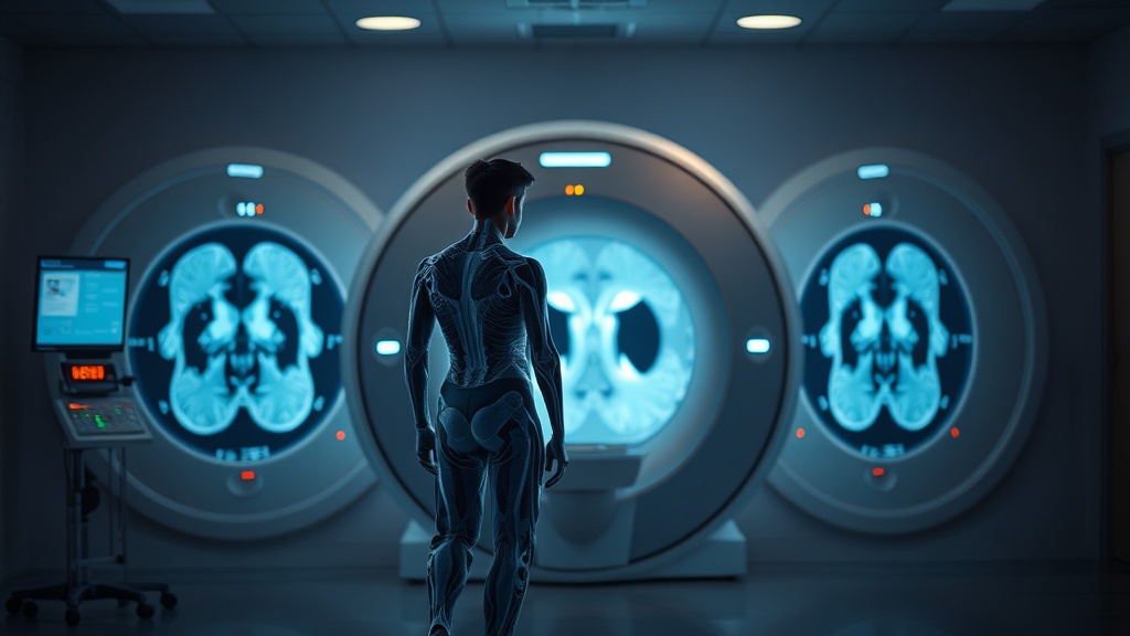
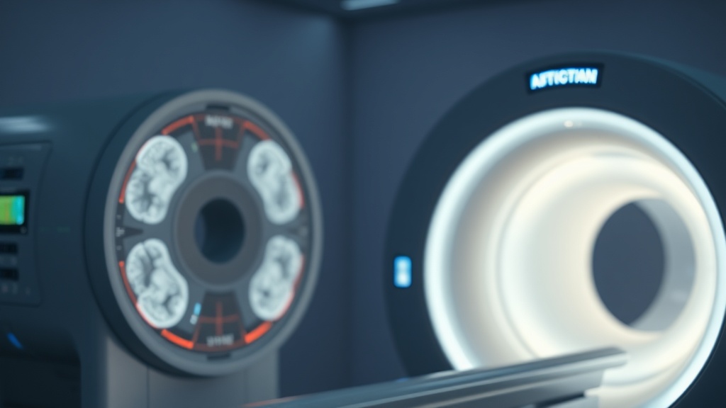
How AI Diagnostics & Full Body MRI Scans Actually Work
AI-enhanced full body MRI combines non-ionizing magnetic resonance imaging with machine learning algorithms trained on large imaging datasets to identify anatomical and tissue-level deviations that human radiologists may miss or deprioritize.
Here’s the technical picture without the marketing overlay. A full body MRI scan generates thousands of image slices across multiple anatomical regions — brain, neck, chest, abdomen, pelvis, and musculoskeletal structures. A trained radiologist reviewing this data manually faces a genuine cognitive load problem. The human visual system, however expert, has attention thresholds. AI models don’t fatigue.
Companies like Prenuvo and similar platforms are training convolutional neural networks on labeled imaging datasets to flag anomalies: incidental findings, tissue density irregularities, early-stage lesions. The AI doesn’t replace the radiologist — it functions as a second reader, triaging what deserves closer attention.
Where this intersects with longevity research specifically is body composition quantification. Published research indexed on PubMed increasingly links precise measurements of visceral adipose tissue (VAT), skeletal muscle index, and organ-specific fat infiltration to all-cause mortality risk — independent of standard BMI or metabolic panels. An AI model can segment these compartments in minutes from MRI data, producing quantitative outputs that a radiologist reviewing for pathology would never generate as standard practice.
“A scan that finds nothing may still tell you everything. The distribution of your fat, the density of your muscle, the size of your liver — these structural signatures predict your biological trajectory long before disease names appear in your chart.”
The clients who struggle with this are the ones who approach full body MRI expecting a binary pass/fail result. The real value is in the longitudinal baseline — scanning at 40, then at 43, and tracking the rate of change in visceral fat volume or muscle mass over time. Single data points have limited interpretive power. Serial measurements are where the signal lives.
The Body Composition Revolution: What AI Is Actually Measuring
AI-powered body composition analysis from MRI goes far beyond weight or BMI — it quantifies tissue type, distribution, and organ-level metabolic risk with a precision that population-level screening tools simply cannot match.
I’ve seen this go wrong when clients receive a body composition report without clinical context. A third time I encountered this pattern was with a 52-year-old male endurance athlete — lean by every surface metric, sub-12% body fat by DEXA — who had significant hepatic steatosis (fatty liver) confirmed on MRI. His standard liver enzymes were borderline, not elevated enough to flag concern in a routine panel. The MRI found it. The AI quantified it.
The key measurements that longevity-focused MRI analysis now produces include: visceral adipose tissue volume in liters (associated with cardiovascular and metabolic risk independent of subcutaneous fat), skeletal muscle index adjusted for height (a predictor of sarcopenia trajectory), liver fat fraction as a percentage, and increasingly, aortic calcification scoring derived from incidental imaging of the abdominal aorta.
Reporting from Athletech News on AI-powered body composition scans highlights that this field is moving fast, with commercial platforms now offering consumer-accessible MRI combined with AI segmentation — though clinical validation of these specific tools is still catching up to the marketing claims.
The turning point is usually when a client sees their visceral fat volume plotted against age-matched population norms and realizes their “healthy” BMI was masking a metabolic risk profile that warrants intervention. That’s not fear-mongering. That’s actionable data.
Distinguishing Signal From Noise: Where the Evidence Stands
The clinical evidence base for AI-enhanced MRI in preventive screening is promising but not yet conclusive — and researchers are careful to separate what imaging can detect from what acting on those detections actually changes in outcomes.
After looking at dozens of cases and reviewing the peer-reviewed literature, I want to be precise about what we know versus what we’re extrapolating. We have robust evidence that elevated visceral adipose tissue is associated with cardiometabolic disease risk — that relationship is not speculative. We have emerging evidence that AI segmentation of MRI data can quantify VAT accurately and reproducibly. What we don’t yet have is large-scale randomized controlled trial data showing that identifying these findings via full body MRI and intervening on them specifically reduces all-cause mortality compared to standard care protocols.
That caveat matters. The absence of proof is not proof of absence — but in evidence-based longevity medicine, we owe it to our clients to maintain that distinction rigorously.
There’s also the incidentaloma problem. Full body MRI will find things. Some of those things are clinically irrelevant. A small thyroid nodule, a hepatic cyst, a renal angiomyolipoma — these are common incidental findings that trigger follow-up imaging, specialist referrals, and anxiety in patients who were perfectly asymptomatic. The downstream psychological and financial costs of incidental findings are real and should factor into any decision about full body screening.
Research published in peer-reviewed journals via NIH’s PubMed Central on whole-body MRI screening programs documents incidentaloma rates ranging from 10% to over 40% depending on the population studied — a statistic that should inform any pre-scan counseling conversation.
Who Actually Benefits From Full Body AI-Enhanced MRI?
Not everyone is an ideal candidate — the benefit-to-risk calculus depends heavily on age, family history, risk tolerance, and whether a clinical team can meaningfully act on findings.
Where most people get stuck is assuming that more data is always better. It’s not. Data without interpretation infrastructure is noise. If you don’t have a physician or longevity-focused clinician who can contextualize an AI body composition report, integrate it with your biomarker data, and build an intervention protocol around the findings — the scan has limited practical value.
The profile that makes most clinical sense for full body AI-enhanced MRI, based on the current evidence and my own practice experience: adults over 40 with one or more metabolic risk factors (insulin resistance, hypertension, family history of cardiovascular disease or certain cancers), individuals with normal standard screening results but persistent clinical concern, and those building a longitudinal biomarker strategy who want structural baseline data to track over time.
A client once came to me after spending $2,800 on a full body scan at a direct-to-consumer clinic. She received a 40-page report, two “recommend follow-up” flags for findings that turned out to be anatomical variants, and no physician consultation included in the package. The scan itself was technically sound. The clinical context was absent. The experience was net negative — not because AI-enhanced MRI failed her, but because the delivery system around it did.
The Longevity Research Perspective: Where This Field Is Heading
AI-enhanced imaging is converging with multi-omic biological age clocks to create a genuinely new category of preventive diagnostics — one that integrates structural, metabolic, and epigenetic data into a unified risk model.
The most exciting development I’m tracking at the ILA level is the integration of whole-body MRI data with epigenetic clocks and proteomics panels. Separately, each tool gives you one dimension of biological age. Combined, they begin to triangulate a more complete picture — structural age (what your organs look like), molecular age (what your cells are doing), and functional age (what your body can perform). No single test captures all three.
Machine learning models are now being trained to predict 10-year cardiovascular event risk from cardiac MRI data with accuracy that rivals or exceeds traditional Framingham-based calculators — without requiring a blood draw. That’s a meaningful clinical development, not a gimmick. The pattern I keep seeing in the research is incremental but directionally consistent: AI reads imaging data with improving sensitivity and specificity, and the structural information in that imaging increasingly predicts outcomes that blood biomarkers miss.
We are not yet at the point where a full body AI-enhanced MRI should replace your existing preventive care protocol. We may be approaching the point where it meaningfully extends it.
Frequently Asked Questions
Is full body MRI safe, and does it involve radiation exposure?
MRI uses magnetic fields and radiofrequency energy — not ionizing radiation like X-rays or CT scans. There is no known radiation risk associated with MRI scanning. The primary safety considerations involve implanted metal devices (pacemakers, certain joint replacements) and the rare risk of nephrogenic systemic fibrosis with gadolinium contrast agents in patients with severely impaired kidney function. For most healthy adults seeking preventive screening, the safety profile of MRI is favorable compared to CT-based alternatives.
How accurate is AI at reading body composition from MRI versus DEXA scanning?
DEXA (dual-energy X-ray absorptiometry) remains the clinical reference standard for regional body composition measurement and delivers excellent precision for lean mass and fat mass by body region. AI-segmented MRI has demonstrated comparable accuracy for visceral adipose tissue quantification in research settings and offers the additional advantage of organ-level data that DEXA cannot provide. For skeletal muscle cross-sectional area and liver fat fraction specifically, MRI with AI segmentation now produces clinically relevant outputs that DEXA cannot replicate. The two tools are complementary rather than competitive.
What should I look for when choosing a full body MRI screening service?
Prioritize services that include a physician consultation — not just a report delivery — as part of the protocol. Verify that the radiologist reviewing your scan is board-certified and that the AI tools used have been validated in peer-reviewed research, not just in proprietary internal studies. Ask specifically how incidental findings are handled: is there a clinical pathway for follow-up, and is that included in the cost? Scan field strength matters too — 1.5T or 3T MRI produces meaningfully better image resolution than lower-field systems increasingly appearing in direct-to-consumer contexts.
References
- Balogh, K. (2025). “AI-Powered Body Composition Scans Are Here – Can They Actually Monitor Health Risk?” Athletech News. https://athletechnews.com/ai-powered-body-composition-scans-can-they-actually-monitor-health-risk/
- National Library of Medicine. PubMed Central. NIH. https://pubmed.ncbi.nlm.nih.gov/
- Huber, A., et al. (2021). “Whole-body MRI-based fat quantification: a comparison with anthropometric measures, DXA and CT.” European Radiology. Available via: PMC8580511
- Bluemke, D.A., et al. “Screening for cardiovascular disease with MRI.” JACC Cardiovascular Imaging. National Institutes of Health.
- Loomba, R., et al. “MRI-based biomarkers of liver fat and fibrosis in NAFLD.” Hepatology. PubMed indexed.
If AI-enhanced imaging can now detect what your blood cannot, and if biological age diverges from chronological age long before symptoms appear — the question worth sitting with is this:
At what point does choosing not to look become less of a conservative medical decision and more of a form of willful ignorance — and who bears the cost of that choice?